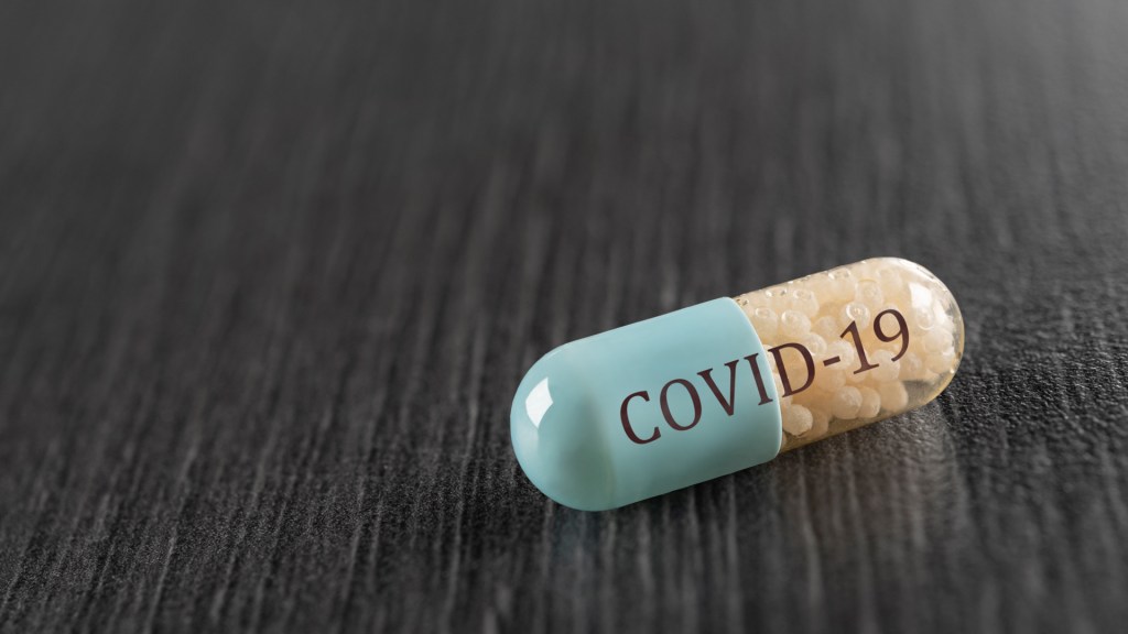
Korean researchers have screened 48 FDA-approved drugs against SARS-CoV-2, and found that two, that are already FDA-approved for other illnesses, seem promising. The FDA approval for other uses would greatly reduce the time needed to gain FDA approval of use in COVID-19. The research is published in Antimicrobial Agents and Chemotherapy, a journal of the American Society for Microbiology. The investigators tested the drugs in Vero cells, a cell line developed from kidney cells of the African Green Monkey, which are commonly used to grow viruses for vaccine production. An anti-helminthic drug called niclosamide demonstrated “very potent” antiviral activity against SARS-CoV-2, according to coauthors Sangeun Jeon, Meehyun Ko, and their collaborators, of the Zoonotic Virus Laboratory, Institut Pasteur Korea, Seongnam, Korea. “Not surprisingly, its broad-spectrum antiviral effect has been well documented in the literature, including antiviral properties against SARS- and MERS-CoV,” they write.
A downside of niclosamide is low absorption, which undercuts the drug’s power by reducing the dose that reaches the target tissue. However, “Further development or drug formulation could enable an effective delivery of this drug to the target tissue,” according to the report. Despite substantially lower antiviral potency, ciclesonide, an inhaled corticosteroid used to treat asthma and allergic rhinitis, also showed promise against SARS-CoV-2. Intriguingly, the investigators note that a study published earlier this year ( by Matsuyama et al.) a treatment report of 3 patients infected by SARS-CoV-2, demonstrated antiviral activity and revealed the drug’s molecular target to be a viral protein called Nsp15. “With its proven anti-inflammatory activity, ciclesonide may represent as a potent drug which can manifest [the] dual roles [of antiviral and anti-inflammatory] for the control of SARS-CoV-2 infection,” the investigators conclude. The anti-inflammatory activity might play a critical role in dampening or preventing the cytokine storms, an immune inflammatory overreaction that can kill COVID-19 patients.
With great pride and honour, we would like to have your gracious presence and to be a part of and support the our upcoming ” 13th International Conference on Pharmaceutical Chemistry (Webinar- Online conference) during May 22, 2020 at Paris, France (Time Zone) due to COVID-19 Pandemic situation (All countries are band to travelling to stop spreading this COVID-2019 Pandemic).
A new observational study from the United States indicates that vitamin D insufficiency may play a significant role in the progression of coronavirus disease (COVID-19). The research titled ‘Vitamin D Insufficiency is Prevalent in Severe COVID-19’ is available on the preprint server medRxiv. A highly transmittable viral infection caused by severe acute respiratory syndrome coronavirus 2 (SARS-CoV-2) is responsible for the current pandemic of potentially fatal COVID-19 disease. The mechanisms underlying the contrasting outcomes of this disease are still unknown. Although 80-85% of patients present with self-limiting disease or do not present with any symptoms, the remaining need significant hospital resources and may potentially collapse the healthcare system, as evidenced by the situation in Italy. The US experience sheds some light on different COVID-19 scenarios and outcomes. Even though they represent only 32% of the US population, African Americans account for 70% of COVID-19 deaths; on the other hand, homeless individuals in Boston shelter were 100% asymptomatic. Risk factors for COVID-19 mortality recognized thus far include male sex, obesity, hypertension, advanced age, and COVID-19 associated coagulopathy. However, vitamin D insufficiency may soon be added to the list of risk factors, according to the researchers from the Louisiana State University Health Sciences Center New Orleans, Tulane School of Medicine, and Texas A&M College of Medicine.
The significance of vitamin D: Vitamin D is a molecule that displays an essential physiological impact and has a pivotal role in modulating both innate and adaptive immune responses. Since the average human diet is not rich in vitamin D of either plant or animal origin, humans depend on endogenous production after ultraviolet B (UVB) exposure. Furthermore, vitamin D-dependent antimicrobial pathways are known to respond to double-stranded RNA, which is readily produced during SARS-CoV-2 viral replication. These pathways subsequently up-regulate the removal of damaged cells (autophagy), as well as various antimicrobial and antiviral peptides. Consequently, vitamin D insufficiency hampers the host’s ability to activate the aforementioned defensive pathways but also been influences lymphocyte and macrophage migration. Since such deranged immune response predisposes the host to viral respiratory infections, is the same valid for COVID-19?
Less vitamin D, worse COVID-19?
To better define the link between the vitamin D insufficiency and COVID-19, the US research group (headed by Dr. Frank H. Lau from the Department of Surgery of the Louisiana State University Health Sciences Center New Orleans) aimed to determine the prevalence of vitamin D insufficiency among our COVID-19 intensive care unit (ICU) patients. The study was performed at a tertiary care academic medical center by retrospectively reviewing medical records of COVID-19 patients and analyzing serum 25-hydroxycholecalciferol (25OHD) levels, which represent the major circulating form of vitamin D. The insufficiency was defined as 25OHD serum levels lower than 30 nanograms per milliliter. The baseline prevalence of vitamin D insufficiency amongst ICU patients was 30-40%; furthermore, 84.6% of ICU patients had the insufficiency (compared to 57.1% of floor patients). The most striking finding was that 100% of ICU patients younger than 75 had vitamin D insufficiency.
The word ‘webinar’ is a blend of ‘web’ and ‘seminar’. A webinar is an event held on the internet which is attended exclusively by an online audience. This distinguishes it from a webcast, which also includes the presence of a physical audience. Other terms used as alternatives for webinar are web event, online seminar, webcast, web lecture and virtual event.
Online participation:
Participants follow webinars via a PC, Mac, tablet or smartphone, and can see and hear the speaker(s) thanks to audio and video feeds. In addition to the video images, PowerPoint slides can be broadcast which run in sync with the rest of the presentation. You can also make use of the screen capture functionality which enables you to show your viewers an application or website.
How does a webinar work?
Before an online seminar can take place, the speaker must first set up an appointment and inform the desired participants about it. Usually the number of participants is limited, so people that want to take part in the webinar must register. The provider then sends a confirmation e-mail to all selected users, which contains further instructions along with the necessary login data and/or access link. It is also common practice to send a reminder message just before the web conference starts. When the webinar begins, participants connect to the web conference room, which requires a standard internet browser. In rare cases, however, it may also be necessary to install a specific client application and use it for the webinar session.
Once you’re connected to the webinar room, you can follow the live broadcast. Typically, you can switch between several windows that present different contents or perform different functions. Normally, there’s the main image, which shows presentation slides or the speaker’s screen for example, and there’s also a slightly smaller window in which the speaker can be seen live. Additional windows may include a list of attendees, a chat feature, or allow access to shared files. If it is a recording, live stream and communication elements are obviously not part of the webinar
The features of online seminars at a glance:
Real-time audio communication via VoIP
Text communication via chat
Uncomplicated presentation of slideshows or screen content
Video streaming
Sharing and downloading of additional material
Practical possibility of recording complete lectures and sharing or viewing them afterwards
Creating and making surveys or quizzes available
Webinar: advantages:
Even if the contents could theoretically be the same, online seminars differ from conventional face-to-face seminars. They offer some advantages compared to classical lectures, but they also come with some disadvantages: the webinar format scores particularly well when it comes to not being dependent on a certain location. This saves time and money for both the speaker and the participants, since they don’t need to travel to the seminar location. Depending on the context of the webinar, anonymous participation is possible in many cases since you only have to enter a valid e-mail address.
Advantages:
Saves costs through no longer having to travel to and from the hotel, overnight stay, room booking, etc.
Simple and automated registration
Easy to exchange information before, during, and after the event
Anonymous participation is possible
In theory, there is no limit to the number of participants (in practice, the maximum number of participants depends on the technical conditions)
Easy to evaluate and store presented content
Join us and gain wonderful experience at Webinar on Pharmaceutical Chemistry during May 22, 2020
“The FDA recognizes that during the COVID-19 public health emergency, the completion of some REMS-required laboratory testing or imaging studies may be difficult because patients suspected of having COVID-19 may be self-isolating and/or subject to quarantine,” said Amy Abernethy, MD, PhD, principal deputy commissioner of the FDA, in a press release. “Under these circumstances, undergoing testing or imaging studies in order to obtain a drug that is subject to a REMS can put patients and others at risk for transmission of the coronavirus. We will continue to work with sponsors to ensure that patients have appropriate access to the medications they need.”
According to the letter, health care providers prescribing and/or dispensing drugs who are subject to REMS with laboratory testing or imagining requirements should consider whether there are compelling reasons not to complete these tests or studies during this public health emergency and use their best medical judgement in weighing the benefits and risks of continuing treatment in the absence of laboratory testing and imaging studies. The FDA does not intend to take action against sponsors and others for the duration of the public health emergency for failing to adhere to REMS requirements for certain laboratory testing or imaging studies.
Additionally, the FDA may require REMS for certain drugs if the agency determines that it is necessary to ensure that the benefits of the drug outweigh its risks. Generally, REMS may include a medication guide, a patient package insert, a communication plan, and certain packaging and safe disposal technologies for drugs that pose a serious risk of abuse or overdose. The agency may also require certain elements to assure safe use (ETASU) as part of REMS for a drug. ETASU are medical interventions or other actions health care professionals need to execute prior to prescribing or dispensing the drug to the patient, such as a requirement to undergo monthly laboratory testing. Some actions may also be required in order for the patient to continue on treatment.
The policy outlined in the guidance will be in effect for the duration of the public health emergency.
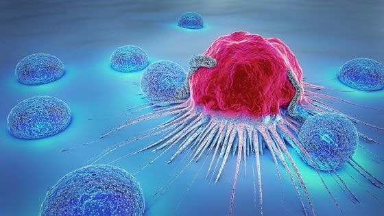
A drug that is already clinically available for the treatment of nausea and psychosis, called prochlorperazine (PCZ), inhibits the internalization of receptors on the surface of tumor cells, thereby increasing the ability of anticancer antibodies to bind to the receptors and mount more effective immune responses. PCZ enhanced the ability of anticancer antibodies to reduce tumor growth in mice, by temporarily increasing the clustering of a receptor targeted by anticancer antibodies on the surface of tumor cells. Temporary treatment of cancer patients with PCZ similarly resulted in receptor clustering on tumor cells inside of patients. The work appears March 5 in the journal Cell.
“This represents a potentially disruptive change in clinical approaches to improve therapeutic targeting as the opportunity of temporarily moving drug targets within patient tumor cells becomes possible,” says senior author Fiona Simpson, a cancer researcher at the University of Queensland. In addition to treating nausea and psychosis, PCZ is currently used in laboratory studies as an inhibitor of endocytosis, a process by which substances are brought into the cell. The temporary inhibition of endocytosis via molecules such as PCZ has been proposed for numerous clinical applications, including control of viral infection, epilepsy, and chronic kidney disease. To help move this approach closer to clinical translation, Simpson and her team recently developed an imaging method to reliably measure endocytosis in humans. When they analyzed tumor samples from patients with squamous cell carcinoma, they found that a decrease in endocytosis of a protein called epidermal growth factor receptor (EGFR) was associated with better clinical responses to the cancer drug cetuximab, an antibody that binds to and inhibits EGFR. “We found that if we stopped the drug targets getting inside the cells by inhibiting the uptake process, called endocytosis, we got a really big immune response against the tumors in mice,” Simpson says.
“This led to the concept that temporarily halting endocytosis of receptors that are the targets of monoclonal antibody therapies may be used to improve clinical outcomes,” Simpson says. Their findings suggested that endocytosis inhibition in humans, which increases the clustering of EGFR on the surface of tumor cells, thereby increasing the availability of the receptor for binding to the antibody, should lead to improved outcomes for patients treated with certain therapeutic antibodies. Exploring this idea in the new study with a different therapeutic antibody, Simpson and her team showed that PCZ improved the immune responses of tumor cells to cetuximab. In mice with cancer, combination therapy with PCZ and cetuximab reversed resistance to cetuximab therapy and inhibited tumor growth more than either treatment alone. Similarly, combination therapy with PCZ and avelumab — an anticancer antibody that inhibits a protein called programmed death ligand 1 (PD-L1) — inhibited tumor growth in mice more than either treatment alone. “These findings highlight the crucial role of availability and persistence of the molecule targeted by the therapeutic antibody at the cell surface of tumor cells,” Simpson says. To begin to test the safety of this approach in humans, Simpson and her team carried out an open-label, uncontrolled, single-center proof-of-concept study in which a single dose of PCZ was intravenously administered to patients with squamous cell carcinoma of the head and neck. Tumor biopsies were taken before and after PCZ treatment. Analysis of results from four patients showed that PCZ increased cetuximab binding sites and the clustering of EGFR on the surface of tumor cells. “As far as we can tell, this was the first verified manipulation of endocytosis in humans,” Simpson says.
The makeup of our microbiomes — the unique communities of bacteria, viruses and other microbes that live in and on us — have been linked, with varying degrees of evidence, to everything from inflammatory bowel disease to athletic performance.
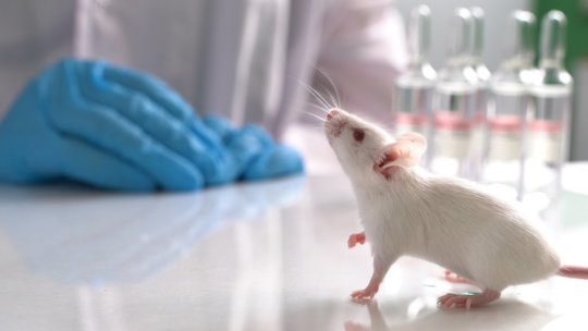
But exactly how could such tiny organisms have such immense influences on a person? : University of California San Diego researchers have created the first-ever map of all the molecules in every organ of a mouse and the ways in which they are modified by microbes. In one surprising example, they discovered that microbes control the structure of bile acids in both mice and people. The study, published February 26, 2020 in Nature, was led by Pieter Dorrestein, PhD, professor and director of the Collaborative Mass Spectrometry Innovation Center in the Skaggs School of Pharmacy and Pharmaceutical Sciences at UC San Diego, and Robert Quinn, PhD, assistant professor at Michigan State University.
When you change the structure of molecules, such as bile acids, you could change how cells talk to one another and which genes are turned “on” or “off” at a given time, Dorrestein said. And that might have huge consequences for body function and the development of disease. “We hear a lot about how our own human genes influence our health and behaviors, so it may come as a shock to think that we could have molecules in the body that look and act the way they do not because of our genes, but because of another living organism,” Dorrestein said. Mapping molecules and microbes in mice: The team compared germ-free (sterile) mice and mice with normal microbes. They used a laboratory technique called mass spectrometry to characterize the non-living molecules in every mouse organ. They identified as many molecules as possible by comparing them to reference structures in the GNPS database, a crowdsourced mass spectrometry repository developed by Dorrestein and collaborators. They also determined which living microbes co-locate with these molecules by sequencing a specific genetic region that acts as a barcode for bacterial types. In total, they analyzed 768 samples from 96 sites of 29 different organs from four germ-free mice and four mice with normal microbes. The result was a map of all of the molecules found throughout the body of a normal mouse with microbes, and a map of molecules throughout a mouse without microbes. A comparison of the maps revealed that as much as 70 percent of a mouse’s gut chemistry is determined by its gut microbiome. Even in distant organs, such as the uterus or the brain, approximately 20 percent of molecules were different in the mice with gut microbes.
Bacteria modify bile acids: After constructing these maps, the researchers homed in on one particular family of molecules that appeared to be significantly different when microbes were present: bile acids. Bile acids are primarily produced by the mouse or human liver, and they help digest fats and oils. They can also carry messages throughout the body. The team discovered bile acids with previously unknown structures in mice with normal microbiomes, but not in germ-free mice. It’s long been known that host liver enzymes add amino acids to bile acids, specifically the amino acids glycine and taurine. But in mice with normal microbiomes, the team found that bacteria are tagging bile acids with other amino acids — phenylalanine, tyrosine and leucine. “More than 42,000 research papers have been published about bile acids over the course of 170 years,” Quinn said. “And yet these modifications had been overlooked.”
Influence on human health: Curious if the same types of microbe-modified bile acids are found in humans, the researchers used a tool they created, the Mass Spectrometry Search Tool (MASST), to search 1,004 public datasets of samples analyzed with mass spectrometry. They also analyzed by mass spectrometry approximately 3,000 fecal samples submitted to the American Gut Project, a large citizen science effort based at UC San Diego School of Medicine. Here’s what they found: The unique microbial-modified bile acids the researchers saw in mice were also present in up to 25.3 percent of all human samples in the datasets. These novel bile acids were more abundant in infants and patients with inflammatory bowel disease or cystic fibrosis. One way bile acids can deliver messages from the gut to other parts of the body is through specific gut receptors called farnesoid X receptors. Bile acids bind and activate the receptors, which then inhibit genes responsible for making more bile acids. The receptors also help regulate liver triglyceride levels and fluid regulation in the intestines, making them important in liver disease and possibly obesity. Several drugs are currently being developed to treat liver disease by activating farnesoid X receptors. Sure enough, in mice and human cells grown in the lab, Dorrestein, Quinn and team found that the newly discovered, microbe-modified bile acids strongly stimulate farnesoid X receptors, reducing expression of genes responsible for bile acid production in the liver. The study raises many questions about the role microbes might play in driving liver and other diseases, and in influencing the activity of therapeutics, such as drugs that target farnesoid X receptors.
“This study provides a clear example of how microbes can influence the expression of human genes,” Dorrestein said. “What we still don’t know is the downstream consequences this could have, or how we might be able to intervene to improve human health.”
Submit your abstracts under sessions “Advanced Organic Chemistry and Inorganic Chemistry”, “Heterocyclic Chemistry”, “Green Chemistry in Pharma Industry”, Biochemistry & Biopharmaceutics” and “Pharmacognosy & phytochemistry”
Benefits:
All accepted abstracts will be published in the respective Supporting Conferences Journals.
Accepted abstracts will be included in the conference proceedings which are to be distributed at the conference.
Each Accepted abstract will receive a Digital Object Identification Number (DOI) provided by CrossRef.
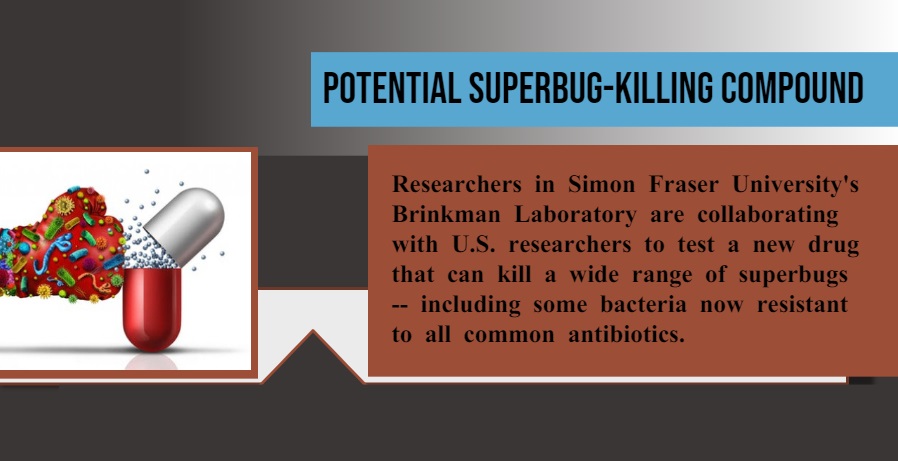
Researchers in Simon Fraser University’s Brinkman Laboratory are collaborating with U.S. researchers to test a new drug that can kill a wide range of superbugs — including some bacteria now resistant to all common antibiotics.
Known as AB569, the drug contains ethylenediaminetetraacetic acid (commonly referred to as EDTA) and acidified nitrite, two inexpensive chemicals that the researchers discovered work together to effectively kill disease-causing bacteria without harming human cells. “We have a growing crisis with antibiotics becoming less and less effective, and treatments are failing; that’s why it’s important to test and develop new drugs and approaches to treat disease-causing bacteria that are highly resistant to existing antibiotics,” says Geoff Winsor, lead database developer at SFU’s Brinkman Lab, which is headed by SFU professor Fiona Brinkman. SFU researchers characterized, at the molecular level, how the chemicals in the AB569 compound were likely working together to kill the notoriously drug-resistant Pseudomonas aeruginosa, using their Pseudomonas Genome Database hosted at SFU, and computer-based analyses of molecular data.
Pseudomonas aeruginosa is a type of bacteria that can cause infections in the lungs (pneumonia), urinary tract, or blood. It is known as the leading cause of morbidity in patients with cystic fibrosis. People who are in hospital or have compromised immune systems are particularly at risk of developing an infection caused by this bacteria. Pseudomonas aeruginosa is categorized by the World Health Organization as a “priority pathogen” of concern. These priority pathogens are highlighted as urgently requiring new treatments, and posing the greatest threat to human health. The top three priority pathogens include highly drug-resistant Acinetobacter baumannii, Pseudomonas aeruginosa, and Enterobacteriaceae. The AB569 compound has been shown to kill these bacteria, plus a wide variety of others, including the notoriously difficult to treat Methicillin-resistant Staphylococcus aureus or MRSA.
“AB569 will go through additional testing because it shows potential as non-toxic topical drug treatment for a wide range of infections,” says Winsor. The lab tests of AB569 showed promising results in treating priority pathogens, plus additional bacteria that cause foodborne illness such as E. coli and Listeria. The AB569 compound was developed by a University of Cincinnati scientist and is now in the first phase of human trials. AB569 has been licensed exclusively to Toronto-based biotechnology firm Arch Biopartners.
We would like to heartily invite you to the upcoming “13th International Conference on Pharmaceutical Chemistry“ which is going to be held Paris, France on May 22-23, 2020. The conference will be focused on theme “Exploring New Challenges, Innovations in Pharmaceutical Chemistry & Drug Discovery”. Opportunities include Oral talks, Poster presentations, workshops, symposiums and Delegate.
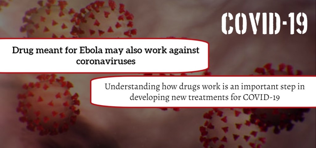
“Even if you know a drug works, it can be a red flag if you don’t know how it works,” said virologist Matthias Götte. “It is reassuring if you know exactly how it works against the target. “We know the drug works against different coronaviruses, like MERS and SARS, and we know the novel coronavirus is very similar to SARS. So I would say I’m cautiously optimistic that the results our team found with remdesivir and MERS will be similar with COVID-19.” The study, published in the Journal of Biological Chemistry this week, is among the first in Canada to discuss the COVID-19 strain. Until now, there has not been a published explanation of why remdesivir may work against coronaviruses, said Götte, who added his study is an important step in answering that question. Developed by Gilead Sciences as a response to the 2014 West African Ebola virus epidemic, remdesivir was first used on a patient with the novel coronavirus earlier this year in the United States.
As reported in the New England Journal of Medicine, the patient was given the drug on the seventh day of illness, and showed marked improvement the following day, with symptoms eventually disappearing altogether. And at a recent press conference in Beijing, the assistant director-general of the World Health Organization, Bruce Alyward, said remdesivir is the only drug available that may have real efficacy against COVID-19. “What our study showed was that remdesivir essentially mimics one of the natural building blocks for RNA synthesis necessary for genome replication of the virus. Enzymes within the virus are synthesizing the viral RNA genome with these building blocks, but they mix up the bits they need with the drug. Once the drug is incorporated into the growing RNA chain, the virus can no longer replicate,”explained Götte. He said the next step is to wait for results from ongoing clinical trials with remdesivir, which are expected by the end of April. Even then, that won’t be the end of the story, he cautioned. “It’s likely we’ll need more than one drug to properly fight emerging diseases like COVID-19, as we have with HIV and hepatitis C virus infections,” Götte said. “Ideally, we will have a couple of drugs because certain strains could be resistant to certain treatments.”
Götte’s study was supported by grants from the Canadian Institutes of Health Research and the Alberta Ministry of Economic Development, Trade and Tourism through the Major Innovation Fund Program and Antimicrobial Resistance — One Health Consortium.
Come and Exploring the Modern Innovation in the field of Pharmaceutical Chemistry at 13th International Conference on Pharmaceutical Chemistry during May 22-23, 2020 at Paris, France. The conference emphasizes the theme “Exploring New Challenges, Innovations in Pharmaceutical Chemistry & Drug Discovery”. It is covers a wide range of critically important sessions, Pharmaceutical Chemistry 2020 is a two-day program which includes thought inspiring keynote presentations, plenary talks, poster presentations, panel discussions, workshops, symposiums, special sessions and career development programs.
The computer model, which can screen more than a hundred million chemical compounds in a matter of days, is designed to pick out potential antibiotics that kill bacteria using different mechanisms than those of existing drugs.
“We wanted to develop a platform that would allow us to harness the power of artificial intelligence to usher in a new age of antibiotic drug discovery,” says James Collins, the Termeer Professor of Medical Engineering and Science in MIT’s Institute for Medical Engineering and Science (IMES) and Department of Biological Engineering. “Our approach revealed this amazing molecule which is arguably one of the more powerful antibiotics that has been discovered.” In their new study, the researchers also identified several other promising antibiotic candidates, which they plan to test further. They believe the model could also be used to design new drugs, based on what it has learned about chemical structures that enable drugs to kill bacteria. “The machine learning model can explore, in silico, large chemical spaces that can be prohibitively expensive for traditional experimental approaches,” says Regina Barzilay, the Delta Electronics Professor of Electrical Engineering and Computer Science in MIT’s Computer Science and Artificial Intelligence Laboratory (CSAIL). Barzilay and Collins, who are faculty co-leads for MIT’s Abdul Latif Jameel Clinic for Machine Learning in Health, are the senior authors of the study, which appears today in Cell. The first author of the paper is Jonathan Stokes, a postdoc at MIT and the Broad Institute of MIT and Harvard.
A new pipeline:
Over the past few decades, very few new antibiotics have been developed, and most of those newly approved antibiotics are slightly different variants of existing drugs. Current methods for screening new antibiotics are often prohibitively costly, require a significant time investment, and are usually limited to a narrow spectrum of chemical diversity. “We’re facing a growing crisis around antibiotic resistance, and this situation is being generated by both an increasing number of pathogens becoming resistant to existing antibiotics, and an anemic pipeline in the biotech and pharmaceutical industries for new antibiotics,” Collins says. To try to find completely novel compounds, he teamed up with Barzilay, Professor Tommi Jaakkola, and their students Kevin Yang, Kyle Swanson, and Wengong Jin, who have previously developed machine-learning computer models that can be trained to analyze the molecular structures of compounds and correlate them with particular traits, such as the ability to kill bacteria. The idea of using predictive computer models for “in silico” screening is not new, but until now, these models were not sufficiently accurate to transform drug discovery. Previously, molecules were represented as vectors reflecting the presence or absence of certain chemical groups. However, the new neural networks can learn these representations automatically, mapping molecules into continuous vectors which are subsequently used to predict their properties.
In this case, the researchers designed their model to look for chemical features that make molecules effective at killing E. coli. To do so, they trained the model on about 2,500 molecules, including about 1,700 FDA-approved drugs and a set of 800 natural products with diverse structures and a wide range of bioactivities. Once the model was trained, the researchers tested it on the Broad Institute’s Drug Repurposing Hub, a library of about 6,000 compounds. The model picked out one molecule that was predicted to have strong antibacterial activity and had a chemical structure different from any existing antibiotics. Using a different machine-learning model, the researchers also showed that this molecule would likely have low toxicity to human cells. This molecule, which the researchers decided to call halicin, after the fictional artificial intelligence system from “2001: A Space Odyssey,” has been previously investigated as possible diabetes drug. The researchers tested it against dozens of bacterial strains isolated from patients and grown in lab dishes, and found that it was able to kill many that are resistant to treatment, including Clostridium difficile, Acinetobacter baumannii, and Mycobacterium tuberculosis. The drug worked against every species that they tested, with the exception of Pseudomonas aeruginosa, a difficult-to-treat lung pathogen. To test halicin’s effectiveness in living animals, the researchers used it to treat mice infected with A. baumannii, a bacterium that has infected many U.S. soldiers stationed in Iraq and Afghanistan. The strain of A. baumannii that they used is resistant to all known antibiotics, but application of a halicin-containing ointment completely cleared the infections within 24 hours.
Preliminary studies suggest that halicin kills bacteria by disrupting their ability to maintain an electrochemical gradient across their cell membranes. This gradient is necessary, among other functions, to produce ATP (molecules that cells use to store energy), so if the gradient breaks down, the cells die. This type of killing mechanism could be difficult for bacteria to develop resistance to, the researchers say. “When you’re dealing with a molecule that likely associates with membrane components, a cell can’t necessarily acquire a single mutation or a couple of mutations to change the chemistry of the outer membrane. Mutations like that tend to be far more complex to acquire evolutionarily,” Stokes says. In this study, the researchers found that E. coli did not develop any resistance to halicin during a 30-day treatment period. In contrast, the bacteria started to develop resistance to the antibiotic ciprofloxacin within one to three days, and after 30 days, the bacteria were about 200 times more resistant to ciprofloxacin than they were at the beginning of the experiment. The researchers plan to pursue further studies of halicin, working with a pharmaceutical company or nonprofit organization, in hopes of developing it for use in humans.
Optimized molecules :
After identifying halicin, the researchers also used their model to screen more than 100 million molecules selected from the ZINC15 database, an online collection of about 1.5 billion chemical compounds. This screen, which took only three days, identified 23 candidates that were structurally dissimilar from existing antibiotics and predicted to be nontoxic to human cells. In laboratory tests against five species of bacteria, the researchers found that eight of the molecules showed antibacterial activity, and two were particularly powerful. The researchers now plan to test these molecules further, and also to screen more of the ZINC15 database. The researchers also plan to use their model to design new antibiotics and to optimize existing molecules. For example, they could train the model to add features that would make a particular antibiotic target only certain bacteria, preventing it from killing beneficial bacteria in a patient’s digestive tract.
The newly-found corbomycin and the lesser-known complestatin have a never-before-seen way to kill bacteria, which is achieved by blocking the function of the bacterial cell wall. The discovery comes from a family of antibiotics called glycopeptides that are produced by soil bacteria. The researchers also demonstrated in mice that these new antibiotics can block infections caused by the drug resistant Staphylococcus aureus which is a group of bacteria that can cause many serious infections.
The findings were published in Nature today.
“Bacteria have a wall around the outside of their cells that gives them shape and is a source of strength,” said study first author Beth Culp, a PhD candidate in biochemistry and biomedical sciences at McMaster.
“Antibiotics like penicillin kill bacteria by preventing building of the wall, but the antibiotics that we found actually work by doing the opposite — they prevent the wall from being broken down. This is critical for cell to divide.
“In order for a cell to grow, it has to divide and expand. If you completely block the breakdown of the wall, it is like it is trapped in a prison, and can’t expand or grow.”
Looking at the family tree of known members of the glycopeptides, researchers studied the genes of those lacking known resistance mechanisms, with the idea they might be an antibiotic demonstrating a different way to attack bacteria.
“We hypothesized that if the genes that made these antibiotics were different, maybe the way they killed the bacteria was also different,” said Culp.
The group confirmed that the bacterial wall was the site of action of these new antibiotics using cell imaging techniques in collaboration with Yves Brun and his team from the Université de Montréal.
Culp said: “This approach can be applied to other antibiotics and help us discover new ones with different mechanisms of action. We found one completely new antibiotic in this study, but since then, we’ve found a few others in the same family that have this same new mechanism.”
The team is led by professor Gerry Wright of the David Braley Centre for Antibiotic Discovery within the Michael G. DeGroote Institute for Infectious Disease Research at McMaster.
The research was funded by the Canadian Institutes of Health Research and the Ontario Research Fund.